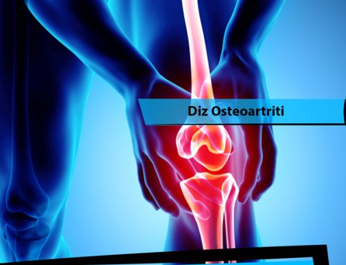
Knee Anatomy
The knee is a hinge-shaped joint between the thigh bone (femur) and the tibia. The joint has two sections: inner and outer. The joint is protected from the front by the kneecap (patella). There are actually 3 joints in the knee. All bone surfaces within the joint are covered with articular cartilage. The load-bearing cartilage surfaces between the femur and tibia are protected and supported by two flexible cartilaginous structures called menisci.
The menisci are a “C” shaped structure with high edges and a thin middle in the shape of a bird’s nest. This structure provides structural harmony between the round femur and the flat tibia, distributes the load across the entire joint surface, absorbs the impacts, and helps stabilize the joint. Ligaments are the main structures that stabilize the knee joint. Ligaments and tendons, which are completely separate structures, should not be confused. Ligaments are fixed structures that attach to the bone at both ends, and they have limited flexibility.
Tendons are structures that attach to the bone at one end and continue with the muscle at the other end, transmitting the movement of the muscle to the bone. The lateral ligaments are located on the inner and outer sides of the knee and prevent the knee from opening to either side. Apart from the external lateral ligament, there is an additional structure called the posterolateral complex, which consists of the ligaments and the popliteus tendon at the back-outer corner of the knee that prevents the knee from opening outward.
The symptoms and treatment of injuries to this structure may be overlooked. The anterior cruciate ligament (ACL) connects the tibia and femur at the exact midpoint. Its function is to limit rotational movements of the knee and prevent the tibia from moving forward. The posterior cruciate ligament (PCL) prevents the tibia from moving backward.
Muscles extend along all of these anatomical structures of the knee and work together to manage movements of the knee such as running and walking. Muscles also support and protect the structures that provide stability. There are two main groups of muscles that manage the knee. The four-headed muscle of the anterior thigh (quadriceps) extends from the pelvic bone along the anterior surface of the thigh, continues by becoming tendons over the kneecap and attaches to the upper-anterior side of the tibia. It performs the movement of straightening the knee (extension).
It also provides balance to the kneecap with separate heads that attach to the upper, inner and outer parts of the kneecap bone. It helps the ACL by restricting the rotational movements of the knee and the PCL by restricting the backward movement of the tibia. There are two hamstring muscles on the back of the thigh that attach to the outside of the tibia and two to the inside. The hamstrings help the ACL by restricting the rotational movement and the forward movement of the tibia. The hamstrings (back thigh muscles) allow the knee to flex (bend).


Leave A Comment